Nanoparticles markedly improve MRI, PET, and fluorescence imaging by enhancing contrast, targeting specific tissues, and enabling real-time visualization. In MRI, they interact with magnetic fields for clearer images, while in PET, they carry radioisotopes for higher sensitivity and better detection of tumors. Fluorescent nanoparticles offer bright, stable signals for cellular imaging. These advances make diagnostics more accurate and personalized. If you want to explore how these technologies are transforming healthcare, keep exploring further.
Key Takeaways
- Nanoparticles serve as contrast agents in MRI, PET, and optical imaging, enhancing image clarity and tissue differentiation.
- Surface chemistry modifications enable targeted delivery and improve biocompatibility for various imaging modalities.
- Superparamagnetic iron oxide nanoparticles (SPIONs) are used in MRI for high sensitivity contrast enhancement.
- Radioactive nanoparticles in PET carry higher radioisotope loads, increasing signal strength and targeting specific tissues.
- Fluorescent nanoparticles like quantum dots provide bright, stable signals for high-resolution cellular and molecular imaging.
Top picks for "nanoparticl imag fluorescence"
Open Amazon search results for this keyword.
As an affiliate, we earn on qualifying purchases.
Enhancing Magnetic Resonance Imaging With Nanoparticles
Nanoparticles have revolutionized magnetic resonance imaging (MRI) by serving as highly effective contrast agents. They improve image quality by enhancing the distinction between healthy and diseased tissues. When introduced into the body, these tiny particles interact with magnetic fields, altering local magnetic properties and producing stronger signals. This results in clearer, more detailed images that help you pinpoint abnormalities with greater accuracy. Unlike traditional contrast agents, nanoparticles can be tailored to target specific tissues or cells, increasing diagnostic precision. Their small size allows them to penetrate tissues more efficiently, providing better spatial resolution. As a result, your ability to detect early-stage diseases improves markedly. Additionally, surface chemistry plays a crucial role in determining nanoparticle biocompatibility and targeting capabilities. Overall, nanoparticle-enhanced MRI offers safer, more sensitive imaging options for better patient outcomes.
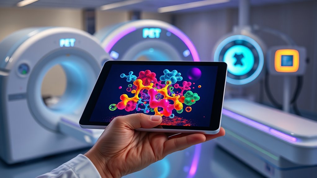
Nanoparticles in Positron Emission Tomography: Improving Sensitivity

By incorporating nanoparticles into positron emission tomography (PET), researchers have markedly enhanced the technique’s sensitivity. Nanoparticles can carry higher loads of radioisotopes, increasing signal strength without raising radiation doses. Their small size allows for improved targeting of specific tissues or tumor sites, reducing background noise and boosting image clarity. Surface modifications enable nanoparticles to bind selectively to biomolecules, further refining detection accuracy. Additionally, nanoparticles can be engineered for prolonged circulation times, granting more time for imaging. Surface modification techniques enable precise targeting and improved biodistribution, enhancing PET imaging capabilities. This combination of increased radioisotope payload and targeted delivery results in sharper, more detailed images, even at lower radiation doses. Overall, integrating nanoparticles into PET significantly advances its ability to detect small lesions and monitor disease progression with greater precision.
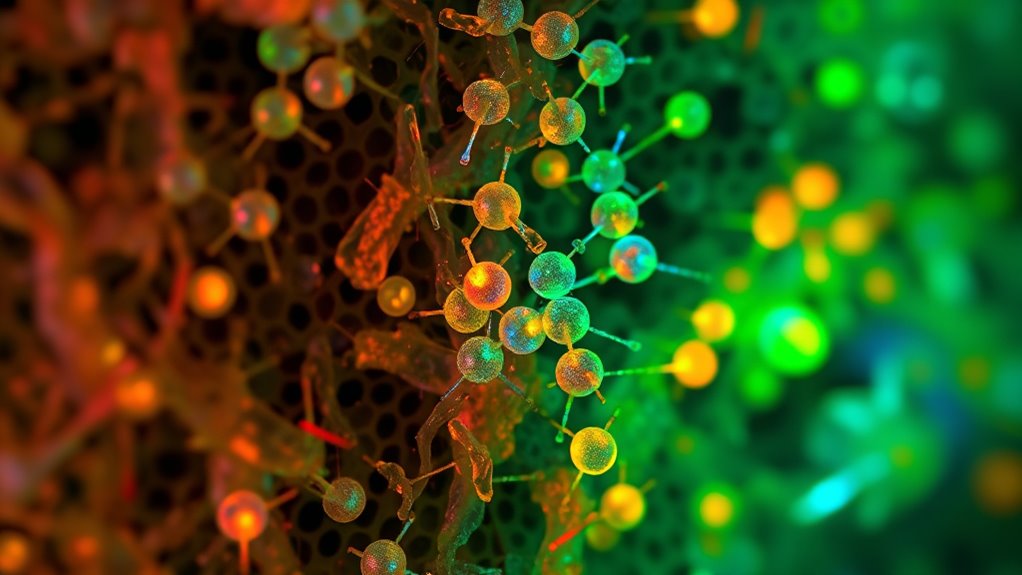
Fluorescent Nanoparticles: Advancing Optical Imaging Techniques

Building on the advancements in nanoparticle-based imaging, fluorescent nanoparticles have emerged as powerful tools for optical imaging. They offer high brightness, photostability, and tunable emission wavelengths, making them ideal for tracking biological processes at the cellular and molecular levels. These nanoparticles can be engineered to target specific tissues or biomarkers, enhancing imaging accuracy. Their ability to emit strong signals under specific light excitation allows for real-time visualization with minimal background interference. You can use them to detect cancer cells, monitor drug delivery, or study cellular interactions. Additionally, their small size enables deep tissue penetration and reduces toxicity concerns. As a result, fluorescent nanoparticles considerably improve the sensitivity and resolution of optical imaging, pushing the boundaries of non-invasive diagnostics. Forsale 100
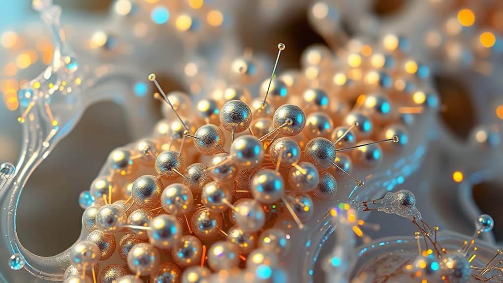
Types of Nanoparticles Used in Medical Imaging

Various types of nanoparticles have been developed for medical imaging, each offering unique advantages tailored to specific diagnostic needs. You might encounter:
Different nanoparticles like SPIONs, quantum dots, and radioactive particles enhance medical imaging with tailored properties.
- Superparamagnetic iron oxide nanoparticles (SPIONs): Ideal for MRI, providing strong contrast and high sensitivity.
- Quantum dots: Fluorescent nanoparticles that enable precise optical imaging with tunable emissions.
- Radioactive nanoparticles: Used in PET scans, delivering targeted signals for metabolic activity and tissue differentiation.
These nanoparticles are engineered with specific properties, such as magnetic, optical, or radioactive features, making them suited for different imaging techniques. Their size, surface chemistry, and stability are tailored to optimize contrast, targeting, and biocompatibility. This variety ensures that you can choose the right nanoparticle for accurate, high-resolution diagnostics. Surface chemistry plays a critical role in enhancing nanoparticle stability and targeting efficiency.
Benefits and Challenges of Using Nanoparticles in Imaging Modalities
Nanoparticles offer significant advantages in medical imaging by enhancing contrast, improving targeting, and enabling early detection of diseases. They can increase sensitivity and specificity, helping you identify abnormalities sooner. Their small size allows them to penetrate tissues more effectively, providing detailed images. However, challenges exist. You must consider potential toxicity and the body’s immune response, which can limit nanoparticle use. Manufacturing consistency and stability are also concerns, as variations can affect imaging quality and safety. Additionally, regulatory approval processes can be lengthy and complex, delaying clinical adoption. Cost is another factor; developing and deploying nanoparticle-based agents may be expensive. Furthermore, understanding how AI Security can support the development and deployment of safe and effective nanoparticle imaging agents is crucial. Balancing these benefits and challenges is essential for advancing nanoparticle applications, ensuring you maximize their potential while addressing safety and practical limitations.
Future Directions and Innovations in Nanoparticle Imaging Technologies

Future innovations in nanoparticle imaging focus on creating multifunctional agents that combine diagnostic and therapeutic capabilities. You’ll see personalized imaging techniques that tailor contrast agents to individual patients, improving accuracy and outcomes. These advancements promise to make imaging more precise, versatile, and patient-specific. Additionally, ongoing research aims to develop advanced storage solutions to enhance the stability and longevity of nanoparticle agents in clinical settings.
Multifunctional Nanoparticles Development
As researchers continue to push the boundaries of imaging technology, developing multifunctional nanoparticles has become a key focus for enhancing diagnostic and therapeutic capabilities. These nanoparticles combine multiple functionalities into a single platform, allowing simultaneous imaging and treatment. You might see designs that integrate targeting ligands, contrast agents, and therapeutic drugs. This integration enables precise delivery, real-time tracking, and multifunctional treatment. Imagine nanoparticles that can identify specific cell types, highlight them in imaging scans, and deliver therapy directly to the target. To achieve this, researchers focus on:
- Combining imaging modalities like MRI and fluorescence within one nanoparticle
- Incorporating targeting molecules for specificity
- Loading therapeutic agents for combined diagnosis and therapy
This approach aims to revolutionize personalized medicine and improve patient outcomes. Gold IRA
Personalized Imaging Techniques
Advancements in nanoparticle design are paving the way for highly personalized imaging solutions that tailor diagnostics to individual patient profiles. You can now develop nanoparticles that target specific biomarkers unique to a person’s disease, enhancing detection accuracy. For example, surface modifications allow nanoparticles to bind selectively to cancer cells or inflamed tissues, providing real-time, precise imaging. This personalization improves early diagnosis, reduces false positives, and guides tailored treatment plans. As you integrate genetic, metabolic, and proteomic data, you can design nanoparticles that respond to specific biological cues, offering dynamic imaging options. Additionally, understanding contrast ratio principles helps optimize image clarity and differentiation in complex biological environments. These innovations enable clinicians to visualize disease progression and treatment response with unprecedented specificity. Ultimately, personalized nanoparticle imaging transforms traditional diagnostics into a precise, patient-centric approach, optimizing outcomes and minimizing invasiveness.
Clinical Applications and Case Studies of Nanoparticle-Based Imaging
You’ll see how nanoparticle-based imaging improves targeted tumor detection, making it easier to identify cancerous tissues. These advanced techniques also boost diagnostic accuracy, helping your healthcare team make better-informed decisions. Examining real case studies highlights the practical benefits and ongoing innovations in this field. Additionally, the use of vibrational energy concepts from the Law of Attraction can be related to maintaining a positive outlook and visualization practices that support health and wellness during treatment.
Targeted Tumor Detection
Targeted tumor detection using nanoparticle-based imaging has revolutionized clinical diagnostics by enabling more precise localization of cancerous tissues. You can now distinguish tumors from healthy tissue with higher accuracy, improving treatment planning. Nanoparticles are tailored to bind specifically to tumor markers, enhancing contrast and visibility during imaging. This specificity reduces false positives and helps identify small or early-stage tumors that traditional methods might miss. The use of spiritual practices such as meditation and prayer can also support patients’ mental health during diagnosis and treatment. You might observe:
- Nanoparticles conjugated with antibodies targeting tumor-specific antigens
- Improved detection of metastases and residual cancer cells
- Enhanced contrast in MRI, PET, or fluorescence imaging for clearer visualization
This approach allows clinicians to make more informed decisions, ultimately leading to better patient outcomes. It’s a significant step toward personalized cancer care, with ongoing advances refining these diagnostic tools further.
Enhanced Diagnostic Accuracy
Nanoparticle-based imaging techniques have considerably improved diagnostic accuracy across various clinical settings. By enhancing contrast and targeting specific tissues, these methods allow you to detect diseases earlier and with greater precision. For example, in cancer diagnosis, nanoparticles improve tumor visibility, reducing false positives and negatives. In cardiovascular imaging, they reveal plaque buildup more clearly, aiding timely intervention. Nanoparticles also enable better differentiation between benign and malignant lesions, guiding treatment decisions. Their ability to deliver high-resolution images benefits many specialties, including neurology and infectious disease. As a result, you can make more informed choices, customize treatments, and improve patient outcomes. Overall, nanoparticle-enhanced imaging elevates the reliability of diagnoses, making clinical assessments faster, more accurate, and more effective.
Frequently Asked Questions
How Do Nanoparticles Cross Biological Barriers for Imaging?
You can facilitate nanoparticles crossing biological barriers through surface modifications like coating them with ligands or polymers that target specific receptors. These modifications help nanoparticles evade immune detection and enhance uptake by cells. Additionally, optimizing particle size and charge allows better navigation through barriers such as the blood-brain barrier. Using external stimuli, like magnetic fields or ultrasound, can also assist in guiding nanoparticles to their desired imaging sites.
Are There Any Long-Term Toxicity Concerns With Nanoparticle-Based Imaging Agents?
You might wonder if nanoparticle-based imaging agents pose long-term risks. While they offer remarkable diagnostic benefits, some concerns exist about their persistence in the body and potential subtle effects over time. Researchers are actively studying their safety profiles, aiming to minimize any lingering impacts. As technology advances, new designs are emerging to balance imaging effectiveness with improved biocompatibility, helping you feel confident in their clinical use.
What Are the Costs Associated With Nanoparticle Production for Medical Imaging?
The costs of producing nanoparticles for medical imaging vary based on materials, complexity, and scale. You’ll find that high-quality, specialized nanoparticles can be expensive due to precise synthesis and purification processes. Large-scale manufacturing may reduce costs over time, but initial R&D, quality control, and regulatory compliance add to expenses. Overall, expect significant investment upfront, but potential long-term savings through improved imaging accuracy and patient outcomes.
Can Nanoparticles Be Targeted to Specific Cell Types or Tissues?
Yes, you can target nanoparticles to specific cell types or tissues. By attaching ligands, antibodies, or peptides to their surface, you enable precise binding to unique markers on target cells. This customization enhances imaging accuracy and reduces side effects. While some worry about complexity or cost, the benefits of improved diagnosis and personalized treatment make targeted nanoparticles a promising tool in medical imaging.
How Do Regulatory Agencies Evaluate Nanoparticle-Based Imaging Agents?
Regulatory agencies evaluate nanoparticle-based imaging agents by reviewing their safety, efficacy, and manufacturing quality. You’ll need to submit detailed data on toxicity, biodistribution, and stability, demonstrating that the particles perform as intended without harmful side effects. Agencies also assess manufacturing processes to guarantee consistency and purity. They may require clinical trial results and post-market surveillance plans to monitor long-term safety, ensuring these advanced agents meet strict standards before approval.
Conclusion
Nanoparticles are revolutionizing medical imaging, offering sharper, more sensitive, and more precise diagnostics. For example, studies show that nanoparticle-enhanced MRI can improve detection accuracy by up to 50%. As technology advances, you’ll see even more innovative applications, making early diagnosis easier and treatment more effective. Embracing these developments means better patient outcomes and a brighter future for personalized medicine. The potential of nanoparticle imaging truly is transforming healthcare as we understand it.