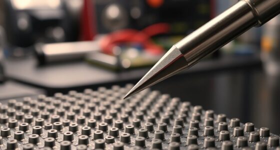

Atomic Force Microscopy (AFM) works by using a tiny cantilever with a sharp tip to scan surfaces at the nanometer scale. As the tip encounters surface features, it experiences forces like van der Waals or electrostatic interactions, causing the cantilever to deflect. A laser reflects off the cantilever into a detector to measure these deflections. By controlling the tip’s position and force, AFM creates high-resolution topographical and surface property maps. If you’re curious, you’ll find more details as you continue to explore.
Key Takeaways
- An AFM uses a cantilever tip that scans a surface to measure nanoscale forces like van der Waals and electrostatic interactions.
- A laser beam reflects off the cantilever into a detector, translating its deflections into surface topography data.
- The microscope operates in modes such as contact or tapping, controlling tip-sample contact to gather different surface information.
- Feedback systems adjust the tip’s position to maintain constant force, ensuring accurate and high-resolution imaging.
- Sensor sensitivity and force regulation enable detailed nanoscale surface characterization through precise deflection measurements.
Atomic Force Microscopy (AFM) is a powerful technique that allows you to visualize surfaces at the nanometer scale. At its core, AFM relies on a tiny probe, called a cantilever tip, that interacts with the sample surface. As you scan the tip across the surface, the forces between the tip and the sample—such as van der Waals, electrostatic, or mechanical forces—cause the cantilever to deflect. This tip sample interaction is fundamental to the technique, as it encodes information about the surface’s topography and properties into the cantilever’s movements.
AFM uses a tiny cantilever tip to scan surfaces and measure forces, revealing nanoscale topography and properties.
The cantilever acts like a sensitive spring, bending slightly in response to the forces it encounters. You measure this deflection using a laser beam reflected off the top of the cantilever into a photodetector. As the tip moves over surface features, the cantilever deflects up or down, and this deflection is directly related to the forces between the tip and the sample. When the tip encounters a bump, groove, or any variation on the surface, the change in cantilever deflection provides a high-resolution map of the surface topography.
During operation, you can choose different modes to extract various types of information. In contact mode, the tip maintains constant contact with the surface, and the cantilever deflection reflects the surface’s topography directly. In tapping mode, the cantilever oscillates near its resonance frequency, intermittently contacting the surface. This mode reduces damage to delicate samples and provides detailed topographical data through changes in oscillation amplitude and phase, still relying heavily on cantilever deflection measurements.
The process involves carefully controlling the tip sample interaction. Too much force risks damaging either the tip or the sample, while too little can reduce resolution. By adjusting the force setpoint and feedback loop, you keep the cantilever deflection within ideal ranges, ensuring accurate data collection. The system continuously adjusts the vertical position of the sample or cantilever to maintain this setpoint during scanning, translating deflections into precise surface images. Additionally, the use of high-resolution sensors enhances the sensitivity and accuracy of the measurements, allowing detailed surface characterization.
Frequently Asked Questions
How Does AFM Compare to Other Microscopy Techniques?
You find AFM stands out because it provides high-resolution surface topography, surpassing many optical microscopes. Unlike electron microscopes, AFM works in various environments, including liquids, making it ideal for nanotechnology advancements. It offers detailed, three-dimensional images at the nanoscale, giving you precise surface information. This makes AFM a versatile tool for exploring materials, biological samples, and nanostructures, often with greater simplicity and less sample preparation.
What Are the Limitations of Atomic Force Microscopy?
Think of AFM as a finely tuned instrument, but it faces hurdles like instrumentation challenges and resolution constraints. You might find it struggles with imaging very soft or sticky samples because the probe can damage or distort them. Its resolution, though impressive, can’t match electron microscopes for atomic detail. These limitations remind you that AFM is powerful but not flawless, requiring careful handling to access its full potential.
Can AFM Be Used for Biological Samples?
Yes, you can use AFM for biological samples, but proper sample preparation is essential. You need to immobilize biological specimens on a suitable substrate and often fix or dehydrate them to prevent damage during imaging. AFM allows high-resolution biological imaging, helping you visualize cell surfaces, proteins, and other biomolecules. Just make certain your samples are compatible with the technique to get accurate, detailed results.
How Is the Data From AFM Interpreted?
You interpret AFM data through force curve analysis, which reveals how the tip interacts with the sample surface, showing properties like elasticity and adhesion. Surface roughness measurement involves analyzing the topographical data to assess surface texture and features. By examining these force curves and topographical images, you can understand material properties at nanoscale resolution, helping you make informed conclusions about sample composition, structure, and surface characteristics.
What Are Common Applications of Atomic Force Microscopy?
Think of AFM as a skilled explorer mapping uncharted territories. You use it to study surface topography, revealing tiny details on materials like cells or nanostructures. It’s also your tool for material characterization, helping you understand properties such as stiffness or conductivity. Whether examining biological samples or nanomaterials, AFM guides you through microscopic worlds, providing insights that shape advancements in medicine, electronics, and materials science.
Conclusion
Now that you know how atomic force microscopy works, you can appreciate its incredible power—it’s like having a microscope strong enough to see individual atoms! With just a tiny tip and some clever technology, you can explore surfaces at an astonishing level of detail. Imagine uncovering secrets hidden at the atomic scale—AFM makes that possible. So, whenever you think about tiny structures, remember, this tool can reveal worlds smaller than you ever imagined.